
SEM vs TEM vs Cryo-EM: Which Electron Microscopy Do You Need?
Quick answer
Choose **SEM** for 3D surface topography of a sample; **TEM** for high-resolution internal ultrastructure of thin sections; and **cryo-EM** to determine near-atomic 3D structures of proteins and complexes in a frozen-hydrated, near-native state. SEM and TEM image morphology; cryo-EM solves molecular structures. In Sweden, AidMix lists electron-microscopy facilities including Lund University and the Umeå Centre for Electron Microscopy (UCEM).
Key takeaways
- SEM = surface/topography, 3D-looking images of the sample exterior
- TEM = electrons pass through thin sections, giving internal ultrastructure at high resolution
- Cryo-EM = frozen-hydrated samples, giving near-atomic 3D structures of biomolecules
- SEM/TEM answer "what does it look like"; cryo-EM answers "what is its 3D structure"
- Sample prep differs hugely — match the method to the question early
"Electron microscopy" isn't one technique. The right choice depends on whether you care about a sample's surface, its internal structure, or the 3D structure of the molecules inside it.
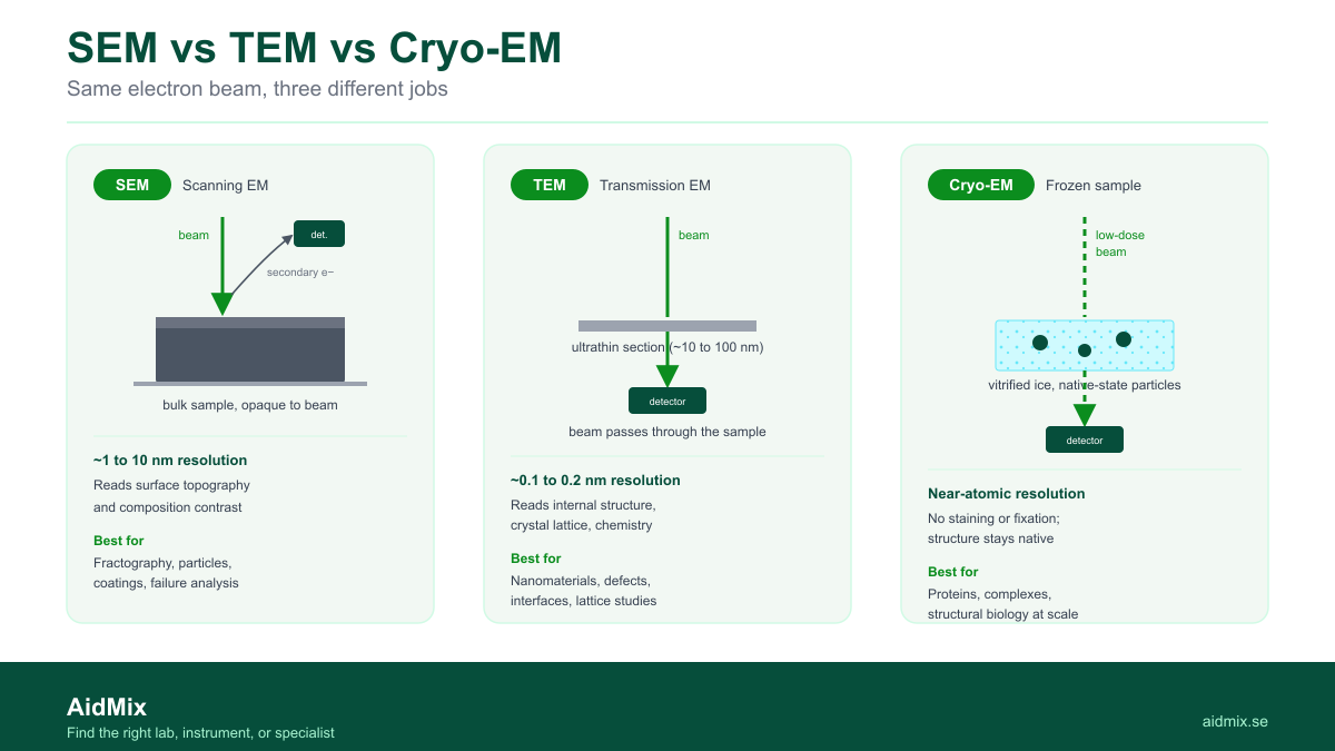
SEM (scanning EM) scans a focused beam across a surface and detects scattered/secondary electrons, producing detailed, almost 3D images of surface topography — great for materials, morphology, and surfaces.
TEM (transmission EM) transmits electrons through an ultra-thin section. It resolves internal ultrastructure at very high resolution — organelles, membranes, nanoparticles, lattice detail.
Cryo-EM flash-freezes samples in vitreous ice and images them near-native, then computationally reconstructs 3D structures of proteins and complexes at near-atomic resolution — without crystallisation. It's a structural biology method, not just imaging.
| | SEM | TEM | Cryo-EM | |---|---|---|---| | Sees | Surface topography | Internal ultrastructure | 3D molecular structure | | Sample | Bulk/coated surface | Ultra-thin section | Frozen-hydrated, near-native | | Resolution | nm-scale surface | Sub-nm internal | Near-atomic (structures) | | Typical use | Materials, morphology | Cell/nanoparticle ultrastructure | Protein/complex structure |
Match to your question: surface → SEM; inside a thin section → TEM; 3D structure of a protein/complex → cryo-EM.
On AidMix: Cryo-EM at Lund University and Titan Krios cryo-TEM at UCEM, Umeå.
Run an EM facility? List it on AidMix for free.
Frequently Asked Questions
Is cryo-EM just a fancy TEM?
Cryo-EM uses transmission electron microscopy on frozen-hydrated samples plus 3D reconstruction to solve molecular structures — a different goal from conventional TEM imaging.
Which gives higher resolution?
For molecular 3D structure, cryo-EM; for internal ultrastructure of sections, TEM; SEM is about surface detail, not internal resolution.
Do I need to coat my sample for SEM?
Non-conductive samples are often sputter-coated; ask the facility about prep for your material.